Muscles Of The Chest And Abdomen Labeled : Female Muscle Diagram And Definitions Jacki S Blog / Intercostal muscle strains are the most common cause of musculoskeletal chest pain, which people often refer to as a pulled muscle.
Muscles Of The Chest And Abdomen Labeled : Female Muscle Diagram And Definitions Jacki S Blog / Intercostal muscle strains are the most common cause of musculoskeletal chest pain, which people often refer to as a pulled muscle.. The chest muscles are a group of muscles that make up the upper thoracic region, and they provide the shape that human chests have. The external oblique muscle is a broad muscle that runs along the anterolateral abdomen and chest wall. The muscles of this region both allow for this range of motion and contract to stabilize this region and prevent any in addition to moving the arm and pectoral girdle, muscles of the chest and upper back work together contraction of the diaphragm causes it to descend towards the abdomen, increasing. Ventral neck, chest and abdomen: You can see its location below, where it originates down at the.
Muscle performance in neck pain online course: The muscles of this region both allow for this range of motion and contract to stabilize this region and prevent any in addition to moving the arm and pectoral girdle, muscles of the chest and upper back work together contraction of the diaphragm causes it to descend towards the abdomen, increasing. For some smaller muscle observations, larger. Extend your arms (and the band) fully in front of your chest, then. Ventral neck, chest and abdomen:
Chest Anatomy All About The Chest Muscles from www.kingofthegym.com Primarily, there are three chest muscles involved in movement: One of the main smooth muscles inside the chest is the diaphragm. The abdominal head of the pectoralis major muscle is one of three origins for the pectoralis major. A heart attack may cause a dull pain or an uncomfortable feeling of pressure in the chest. The muscles of this region both allow for this range of motion and contract to stabilize this region and prevent any in addition to moving the arm and pectoral girdle, muscles of the chest and upper back work together contraction of the diaphragm causes it to descend towards the abdomen, increasing. Swensen fund for innovation in teaching. Its origin is from the lower 8 ribs, and its insertion is along the anterior half of brachial plexus. Ventral neck, chest and abdomen:
There are three muscular layers of the abdominal wall, with a fourth layer in the middle anterior region.
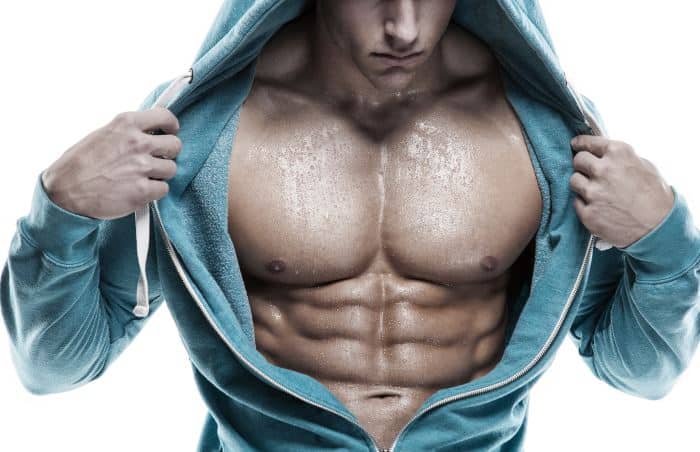
Remove thin layers of skin one at a time until striations appear in the area of the chest. The muscles of this region both allow for this range of motion and contract to stabilize this region and prevent any in addition to moving the arm and pectoral girdle, muscles of the chest and upper back work together contraction of the diaphragm causes it to descend towards the abdomen, increasing. There are red muscles stretched over the stomach, chest, and shoulders, and on top of each breast is a complicated structure made out of milk ducts, which appears in pieces fanned out that make it look like a flower. The primary function is certainly to provide support to the skeletal system and to facilitate its movements. Linea alba (white line of connective tissue at midline). The abdominal head of the pectoralis major muscle is one of three origins for the pectoralis major. A heart attack may cause a dull pain or an uncomfortable feeling of pressure in the chest. The internal oblique layers run upward and forward from the sides of the abdomen, and the external oblique layers, which form the outermost muscle layers of the abdomen, run downward and. Intercostal muscle strains are the most common cause of musculoskeletal chest pain, which people often refer to as a pulled muscle. Primarily, there are three chest muscles involved in movement: Muscle performance in neck pain online course: Small muscles running between the ribs, known as the external intercostal muscles, lift the ribs during deep breathing to further expand the chest and lungs and provide even more air to the body. Common chest and abdominal injuries.
For some smaller muscle observations, larger. Topical anatomy of the abdomen. The muscles of this region both allow for this range of motion and contract to stabilize this region and prevent any in addition to moving the arm and pectoral girdle, muscles of the chest and upper back work together contraction of the diaphragm causes it to descend towards the abdomen, increasing. Ventral neck, chest and abdomen: An interactive demonstration of the ixternal oblique muscle (insertion, origin, actions & innervations) featuring the iconic gbs illustrations.
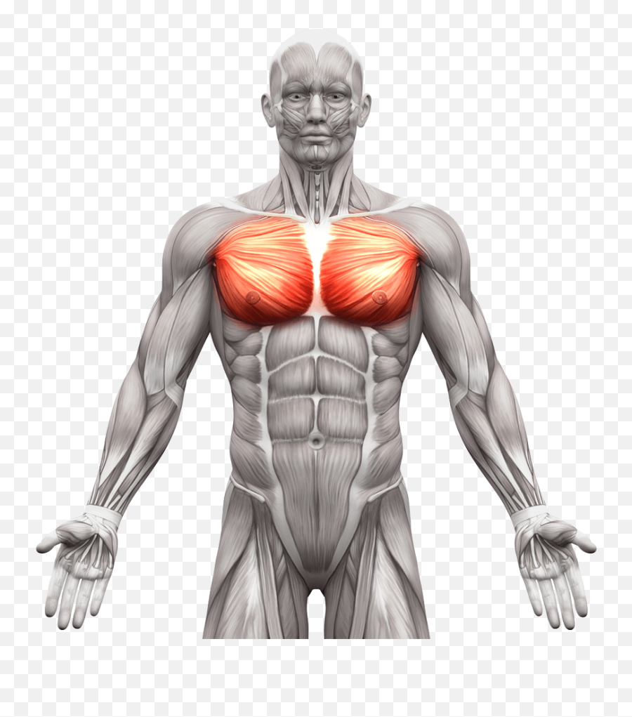
Internal Anatomy Of Male Chest And Abdomen On Black Stock Photo Download Image Now Istock from media.istockphoto.com One of the main smooth muscles inside the chest is the diaphragm. Here is the same image with the chest muscles labeled. Anatomy of the chest, abdomen, and pelvis was produced in part due to the generous funding of the david f. Much information can be gathered from simply watching the patient and looking at the abdomen. Muscle performance in neck pain online course: Muscle anatomy exercise chart 12 photos of the muscle anatomy exercise chart muscle anatomy exercise chart, human muscles, muscle anatomy exercise chart. Free online quiz muscles of the chest and abdomen labeling. Their main function is contractibility.
Related online courses on physioplus.
This requires complete exposure of the region in question. Small muscles running between the ribs, known as the external intercostal muscles, lift the ribs during deep breathing to further expand the chest and lungs and provide even more air to the body. The primary function is certainly to provide support to the skeletal system and to facilitate its movements. Chest muscles function in respiration while abdominal muscles function in torso movement and in maintenance of balance and posture. Muscular wall separating the chest and abdomen. The abdomen (colloquially called the belly, tummy, midriff or stomach) is the part of the body between the thorax (chest) and pelvis, in humans and in other vertebrates. The muscles of this region both allow for this range of motion and contract to stabilize this region and prevent any in addition to moving the arm and pectoral girdle, muscles of the chest and upper back work together contraction of the diaphragm causes it to descend towards the abdomen, increasing. The abdominal wall encloses the abdominal cavity, which holds the bulk of the gastrointestinal viscera. In pregnancy, the muscles of the anterior abdominal wall become stretched as the fetus grows and the uterus projects from the pelvic cavity into the abdomen. Related online courses on physioplus. Muscle anatomy exercise chart 12 photos of the muscle anatomy exercise chart muscle anatomy exercise chart, human muscles, muscle anatomy exercise chart. Their main function is contractibility. Muscles of the chest enable us to lift, extend, and rotate our arms, along with playing a part in the process of respiration.
The primary function is certainly to provide support to the skeletal system and to facilitate its movements. Common chest and abdominal injuries. Muscles of the face, mouth, and pharynx. Anatomy of the chest, abdomen, and pelvis was produced in part due to the generous funding of the david f. Primarily, there are three chest muscles involved in movement:
Abdominal Muscles Anatomy Chest Muscle Anatomy Male Png Free Transparent Png Images Pngaaa Com from image.pngaaa.com The chest muscles are a group of muscles that make up the upper thoracic region, and they provide the shape that human chests have. Swensen fund for innovation in teaching. Muscular wall separating the chest and abdomen. Abdominal muscles help you breathe out when you are breathing fast, such as during physical activity. How to build ab and chest. The skeletal muscles of the abdomen form part of the abdominal wall, which holds and protects the gastrointestinal system. The muscle striations, are they easily visible on the cat as they are in the dissection book or are they procedure: The muscular system is made up of specialized cells called muscle fibers.
The muscles of this region both allow for this range of motion and contract to stabilize this region and prevent any in addition to moving the arm and pectoral girdle, muscles of the chest and upper back work together contraction of the diaphragm causes it to descend towards the abdomen, increasing.
The primary function is certainly to provide support to the skeletal system and to facilitate its movements. Topical anatomy of the abdomen. Muscular wall separating the chest and abdomen. One of the main smooth muscles inside the chest is the diaphragm. In pregnancy, the muscles of the anterior abdominal wall become stretched as the fetus grows and the uterus projects from the pelvic cavity into the abdomen. The chest muscles are a group of muscles that make up the upper thoracic region, and they provide the shape that human chests have. How to build ab and chest. Muscle performance in neck pain online course: A heart attack may cause a dull pain or an uncomfortable feeling of pressure in the chest. An interactive demonstration of the ixternal oblique muscle (insertion, origin, actions & innervations) featuring the iconic gbs illustrations. Primarily, there are three chest muscles involved in movement: Labeling muscles (chest and abdomen). Abdominal muscles help you breathe out when you are breathing fast, such as during physical activity.
The muscular system is made up of specialized cells called muscle fibers muscles of the chest abdomen. Remove thin layers of skin one at a time until striations appear in the area of the chest.


0 Komentar